DEXA

What is a Bone Density (DEXA)?

Bone density scanning, also called dual-energy x-ray absorptiometry (DXA or DEXA) or bone densitometry, is an enhanced form of x-ray technology that is used to measure bone loss. DEXA is today’s established standard for measuring bone mineral density (BMD).

An x-ray (radiograph) is a painless medical test that helps physicians diagnose and treat medical conditions. Radiography involves exposing a part of the body to a small dose of ionizing radiation to produce pictures of the inside of the body. X-rays are the oldest and most frequently used form of medical imaging.

DEXA is most often performed on the lower spine and hips. Portable DEXA devices, including some that use ultrasound waves rather than x-rays, measure the wrist, fingers or heel and are sometimes used for screening purposes.

DEXA

Our Hologic Discovery QDR Bone Density machine is a industry leader. Hologic sets the standard for bone density with this advanced patient friendly machine.

Our Commitment

  • WET Reads/STAT Reads in 2-4 hours
  • Online Film/Report viewing for Physicians
  • Onsite Radiologists
  • Late Hours/Appointments
  • Creatinine Testing Onsite
  • Friendly and Helpful Staff
  • ACR Accredited Machines
  • Certified Technologists
  • Complimentary Film/CD Delivery to Physicians

What are some common uses of the procedure?

DEXA Bone Scan
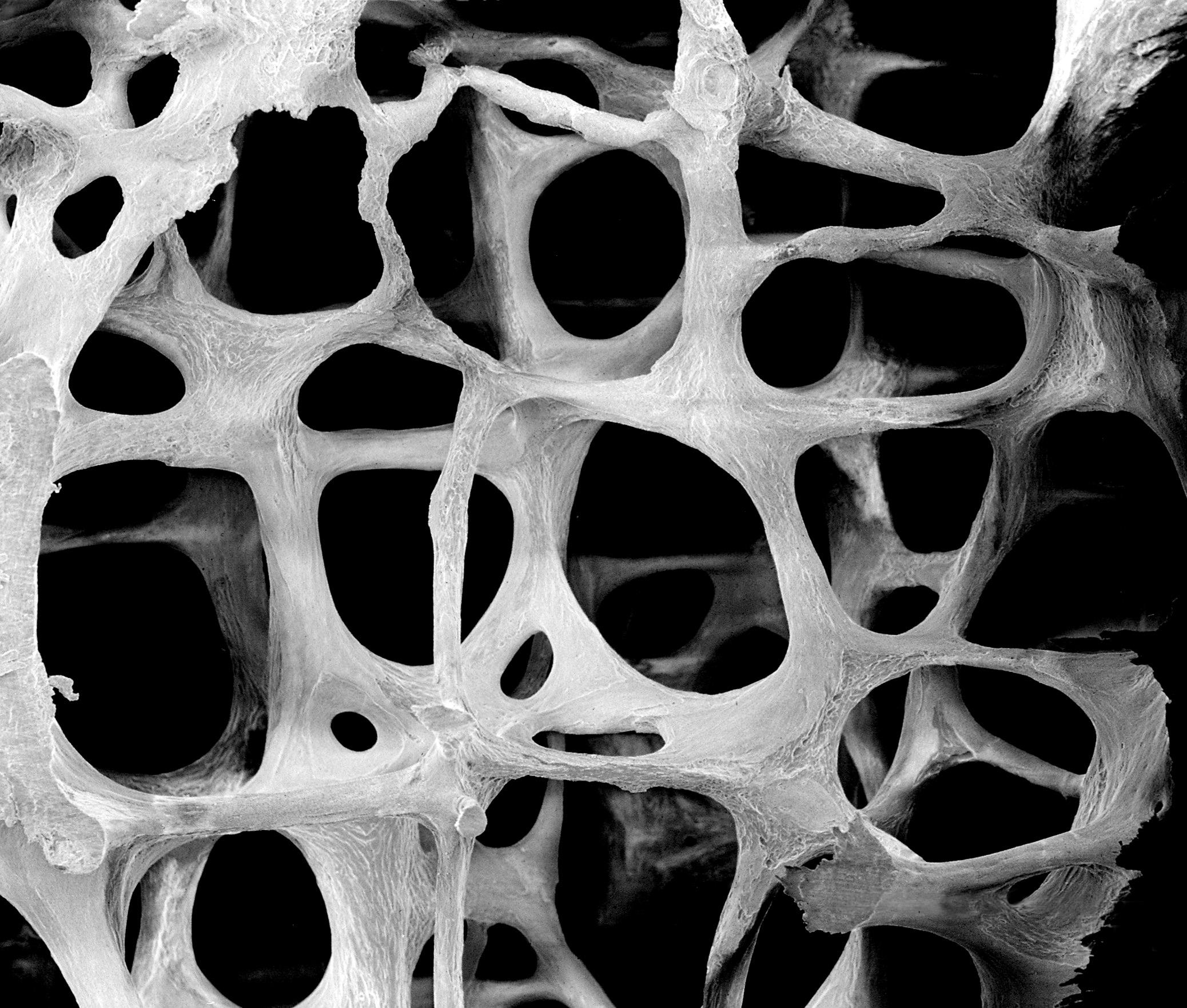
DEXA bone densitometry is most often used to diagnose osteoporosis, a condition that often affects women after menopause but may also be found in men. Osteoporosis involves a gradual loss of calcium, causing the bones to become thinner, more fragile and more likely to break.

DEXA is also effective in tracking the effects of treatment for osteoporosis and other conditions that cause bone loss.

The DEXA test can also assess an individual’s risk for developing fractures.

Bone density testing is strongly recommended if you:

  • Are a post-menopausal woman and not taking estrogen.
  • Have a personal or maternal history of hip fracture or smoking.
  • Are a post-menopausal woman who is tall (over 5 feet 7 inches) or thin (less than 125 pounds).
  • Are a man with clinical conditions associated with bone loss.
  • Use medications that are known to cause bone loss, including corticosteroids such as Prednisone, various anti-seizure medications such as Dilantin and certain barbiturates, or high-dose thyroid replacement drugs.
  • Have type 1 (formerly called juvenile or insulin-dependent) diabetes, liver disease, kidney disease or a family history of osteoporosis.
  • Have high bone turnover, which shows up in the form of excessive collagen in urine samples.
  • Have a thyroid condition, such as hyperthyroidism.
  • Have a parathyroid condition, such as hyperparathyroidism.
  • Have experienced a fracture after only mild trauma.
  • Have had x-ray evidence of vertebral fracture or other signs of osteoporosis.

How does the procedure work?

The DEXA machine sends a thin, invisible beam of low-dose x-rays with two distinct energy peaks through the bones being examined. One peak is absorbed mainly by soft tissue and the other by bone. The soft tissue amount can be subtracted from the total and what remains is a patient’s bone mineral density.

DEXA machines feature special software that compute and display the bone density measurements on a computer monitor.

What will I experience during and after the X-ray procedure?

Bone density tests are a quick and painless procedure.

How is the procedure performed?

This examination is usually done on an outpatient basis. In the Central DEXA examination, which measures bone density in the hip and spine, the patient lies on a padded table. An x-ray generator is located below the patient and an imaging device, or detector, is positioned above.

To assess the spine, the patient’s legs are supported on a padded box to flatten the pelvis and lower (lumbar) spine. To assess the hip, the patient’s foot is placed in a brace that rotates the hip inward. In both cases, the detector is slowly passed over the area, generating images on a computer monitor.

The patient must hold very still and may be asked to keep from breathing for a few seconds while the x-ray picture is taken to reduce the possibility of a blurred image. The technologist will walk behind a wall or into the next room to activate the x-ray machine.

The peripheral DEXA (pDEXA) test is even simpler. The patient’s finger, hand, forearm or foot is placed a small device that obtains a bone density reading within a few minutes.

An additional procedure called Lateral Vertebral Assessment (LVA) is now being done at many centers. LVA is a low-dose x-ray examination of the spine to screen for vertebral fractures that is performed on the DEXA machine.

The LVA test adds only a few minutes to the DEXA procedure.

The DEXA bone density test is usually completed within 10 to 30 minutes, depending on the equipment used and the parts of the body being examined.